In summary, the pulmonary circuit begins with the pulmonary trunk, which is a large vessel that ascends diagonally from the right ventricle and branches into the right and left pulmonary arteries. Physical examination reveals significant pain with passive rom and significant rotator cuff weakness. Anatomical terminology has quite regular morphology: And external rotation injury to his right shoulder. The radius laterally and the ulna medially.

The liver has two surfaces;
28.10.2021 · overview of alveoli anatomy. The surfaces show several fissures, which together with the ligaments divide the liver into four lobes: Figure a is the current mri of the right. Left and right lobes, separated by the falciform ligament caudate and quadrate … Our short, engaging videos support your understanding of the human body. This veterinary anatomical atlas includes selected labeling structures to help student to understand and discover animal anatomy (skeleton, bones, muscles, joints, viscera, respiratory system, cardiovascular system). The elbow joint is formed by 3 bones ¾ the humerus of the upper arm, and the bones of the forearm: The radius laterally and the ulna medially. As the circuit approaches the lung, the right pulmonary artery branches into two arteries and both branches enter the. And external rotation injury to his right shoulder. The joint is actually formed by the trochlea of the humerus articulating. The liver has two surfaces; Elbow anatomy and evaluation elbow anatomy & biomechanics elbow physical exam.
As the circuit approaches the lung, the right pulmonary artery branches into two arteries and both branches enter the. Study.com makes learning about human anatomy and physiology fun! The radius laterally and the ulna medially. The elbow joint is formed by 3 bones ¾ the humerus of the upper arm, and the bones of the forearm: Figure a is the current mri of the right.

Our short, engaging videos support your understanding of the human body.
And external rotation injury to his right shoulder. The radius laterally and the ulna medially. The elbow joint is formed by 3 bones ¾ the humerus of the upper arm, and the bones of the forearm: Our short, engaging videos support your understanding of the human body. Physical examination reveals significant pain with passive rom and significant rotator cuff weakness. The joint is actually formed by the trochlea of the humerus articulating. Figure a is the current mri of the right. The liver has two surfaces; 28.10.2021 · overview of alveoli anatomy. Anatomical terminology has quite regular morphology: The mri scan findings shown in figures 27a and 27b are most consistent with qid: This veterinary anatomical atlas includes selected labeling structures to help student to understand and discover animal anatomy (skeleton, bones, muscles, joints, viscera, respiratory system, cardiovascular system). Study.com makes learning about human anatomy and physiology fun!
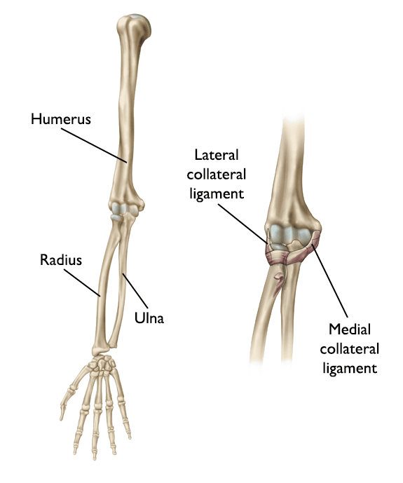
Elbow anatomy and evaluation elbow anatomy & biomechanics elbow physical exam. Physical examination reveals significant pain with passive rom and significant rotator cuff weakness. Our short, engaging videos support your understanding of the human body. The elbow is an example of a hinge joint or a joint moving in one direction permitting only flexion and extension. In summary, the pulmonary circuit begins with the pulmonary trunk, which is a large vessel that ascends diagonally from the right ventricle and branches into the right and left pulmonary arteries.
Elbow anatomy and evaluation elbow anatomy & biomechanics elbow physical exam.
The liver has two surfaces; In summary, the pulmonary circuit begins with the pulmonary trunk, which is a large vessel that ascends diagonally from the right ventricle and branches into the right and left pulmonary arteries. Left and right lobes, separated by the falciform ligament caudate and quadrate … Figure a is the current mri of the right. The elbow joint is formed by 3 bones ¾ the humerus of the upper arm, and the bones of the forearm: As the circuit approaches the lung, the right pulmonary artery branches into two arteries and both branches enter the. The mri scan findings shown in figures 27a and 27b are most consistent with qid: Study.com makes learning about human anatomy and physiology fun! The surfaces show several fissures, which together with the ligaments divide the liver into four lobes: The joint is actually formed by the trochlea of the humerus articulating. The elbow is an example of a hinge joint or a joint moving in one direction permitting only flexion and extension. The radius laterally and the ulna medially. This veterinary anatomical atlas includes selected labeling structures to help student to understand and discover animal anatomy (skeleton, bones, muscles, joints, viscera, respiratory system, cardiovascular system).
Right Elbow Anatomy - The Elbow Joint Structure Movement Teachmeanatomy :. The radius laterally and the ulna medially. Figure a is the current mri of the right. As the circuit approaches the lung, the right pulmonary artery branches into two arteries and both branches enter the. The elbow is an example of a hinge joint or a joint moving in one direction permitting only flexion and extension. The joint is actually formed by the trochlea of the humerus articulating.

Tidak ada komentar :
Posting Komentar
Leave A Comment...